Pesquisa
US Quiz of the Month – Agosto 2024
Case Report
We present the case of a 28-year-old male with past history of non-stricturing, non-penetrating ileocolic Crohn’s disease with associated perianal involvement, currently medicated with Ustekinumab SC, 90mg every 4 weeks. An abdominal CT scan was performed to evaluate the disease activity, which detected a 16mm hypervascular lesion the pancreatic tail, suggestive of a neuroendocrine tumor. The lesion was further characterized by a magnetic resonance cholangiopancreatography (MRCP), without any specific characteristics and suggesting correlation with EUS findings (Fig. 1).
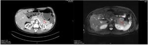
Figure 1. CT and MRI images showing an hipervascular pancreatic lesion (arrow).
During the EUS a 16x9mm homogenous nodular lesion was identified in the pancreatic tail (Fig. 2). The lesion had regular contours and well-defined borders and was hyperechoic in relation to the pancreatic and splenic parenchyma. Elastography predominantly showed a green pattern, with an SR of approximately 2.3. Contrast-enhanced US (CEUS) revealed late contrast hyperenhancement compared to the adjacent pancreatic parenchyma.
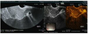
Figure 2. EUS image showing: a) a 13.2×9.0mm solid lesion; b) hypervascular behaviour on CEUS.
What is the most likely diagnosis?
Discussion
A fine-needle biopsy confirmed the diagnosis of accessory spleen. An accessory spleen, or splenunculus, is a congenital anomaly characterized by the presence of ectopic splenic tissue that did not fuse during embryonic development. [1]
The most frequent locations for their development are the splenic hilum, vessels and pancreatic tail.2 Usually they are incidentally found during imaging exams done for other conditions. [2]
Although benign and often clinically insignificant, it is important to recognize them, to avoid unnecessary interventions and to differentiate them from other entities such as pancreatic neuroendocrine tumors or metastatic disease. [1]
In this case, however, the accessory spleen presented with a slightly different ultrasonographic appearance from the normal splenic tissue thus raising the possibility of differential diagnostics and histological confirmation made it possible to rule out other entities with clinical significance.
References
- Trujillo SG, Saleh S, Burkholder R, Shibli F, Shah B. Accessory Spleen: A Rare and Incidental Finding in the Stomach Wall. Cureus. 2022 May 13;14(5):e24977. doi: 10.7759/cureus.24977. PMID: 35698709; PMCID: PMC9188829.
- Vikse J, Sanna B, Henry BM, Taterra D, Sanna S, Pękala PA, Walocha JA, Tomaszewski KA. The prevalence and morphometry of an accessory spleen: A meta-analysis and systematic review of 22,487 patients. Int J Surg. 2017 Sep;45:18-28. doi: 10.1016/j.ijsu.2017.07.045. Epub 2017 Jul 15. PMID: 28716661.
Authors
João Afonso1,2, Pedro Moutinho Ribeiro1,2, Rosa Coelho1,2, Guilherme Macedo1,2.
- Gastroenterology Department, Centro Hospitalar Universitário São João, Porto, Portugal.
- Faculty of Medicine of the University of Porto, Porto, Portugal.